Atrial flutter: from ECG to electroanatomical 3D mapping
Abstract
Overview
Atrial flutter is a common arrhythmia that may cause significant symptoms, including
palpitations, dyspnea, chest pain and even syncope. Frequently it’s possible to diagnose
atrial flutter with a 12-lead surface ECG, looking for distinctive waves in leads II, III, aVF, aVL,
V1,V2. Puech and Waldo developed the first classification of atrial flutter in the 1970s. These authors
divided the arrhythmia into type I and type II. Therefore, in 2001 the European Society of
Cardiology and the North American Society of Pacing and Electrophysiology developed a new
classification of atrial flutter, based not only on the ECG, but also on the electrophysiological
mechanism. New developments in endocardial mapping, including the electroanatomical 3D
mapping system, have greatly expanded our understanding of the mechanism of arrhythmias.
More recently, Scheinman et al, provided an updated classification and nomenclature. The terms
like common, uncommon, typical, reverse typical or atypical flutter are abandoned because they
may generate confusion. The authors worked out a new terminology, which differentiates atrial flutter
only on the basis of electrophysiological mechanism. (Heart International 2006; 3-4: 161-70)
Keywords
Atrial flutter, Catheter ablation, ECG, Electroanatomical mapping
Article Information
Correspondence
Dr. Giuseppe Inama, Department of Cardiology, Ospedale Maggiore, Largo U. Dossena, 2, 26013 Crema – Italy, g.inama@hcrema.it

Trending Topic
As of the publication of this article, the coronavirus disease 2019 (COVID-19) infection has affected over 400 million people around the world and caused over 6 million deaths.1 Although COVID-19 infection predominantly affects the respiratory system, studies have described a wide spectrum of cardiovascular manifestations, including asymptomatic myocardial injury, myocardial infarction and myocarditis.2 Echocardiography is an easily […]
Related Content in Imaging

More than half of acute coronary syndromes (ACS) occur in subjects with no significant coronary stenosis.1 In view of the number of patients dying each year from a heart attack, the question of identifying these patients is of primary importance. ...

Left main Stenosis Stenting Normalizes Wall Shear Stress of Ascending Aorta in Bicuspid Aortic Valve
Bicuspid aortic valve (BAV) represents the most common congenital cardiac anomaly, with a prevalence ranging between 1% and 2% in the general population.1 BAV is known to be associated with dilation and dissection of the ascending aorta, and the significantly higher shear ...

Anomalous coronary arteries originating from the opposite sinus of Valsalva (ACAOS) is one of the most clinically relevant abnormalities among the wide spectrum of coronary artery anomalies.1 Sudden cardiac death has been related to myocardial bridges, ectopic origin of the ...

Introduction: The restriction of activation mapping to the ventricular surface of contemporary mapping systems often leads to failure to correctly identify the true site of origin (SOO) of intramural and/or sub-epicardial Vas and lower procedural success. Electromechanical wave imaging (...

Introduction: Leadless left ventricular (LV) endocardial pacing with the WiSE-CRT System (EBR Systems Inc) is a novel treatment in the field of cardiac resynchronization therapy (CRT). The system was designed to provide lateral wall LV pacing (LVP) in response to ...

Background: The spatial resolution of a mapping catheter is determined by factors such as the quantity of electrodes, their inter-electrode spacing, and the various configurations in which they are utilized. These factors impact the interpretation of wavefront dynamics during cardiac ...

Background: Obesity-related cardiac remodelling may be reflected through electrocardiogram (ECG) changes. Based on this premise, we hypothesised that an artificial intelligence (AI)-ECG model could be trained to predict body mass index (BMI), and that the difference between AI-ECG predicted ...

Athletes represent the fittest individuals in our society, yet paradoxically carry an increased risk of sudden cardiac death (SCD) when compared to sedentary individuals with the same cardiac disease.1,2 Two recent studies have provided a realistic prevalence of young SCD ...

Sudden cardiac death arises from the unexpected stopping of the heart and is associated with up to a half of cardiovascular deaths. We discuss with Professor Martin Borggrefe how to identify patient populations at risk of sudden cardiac death (...

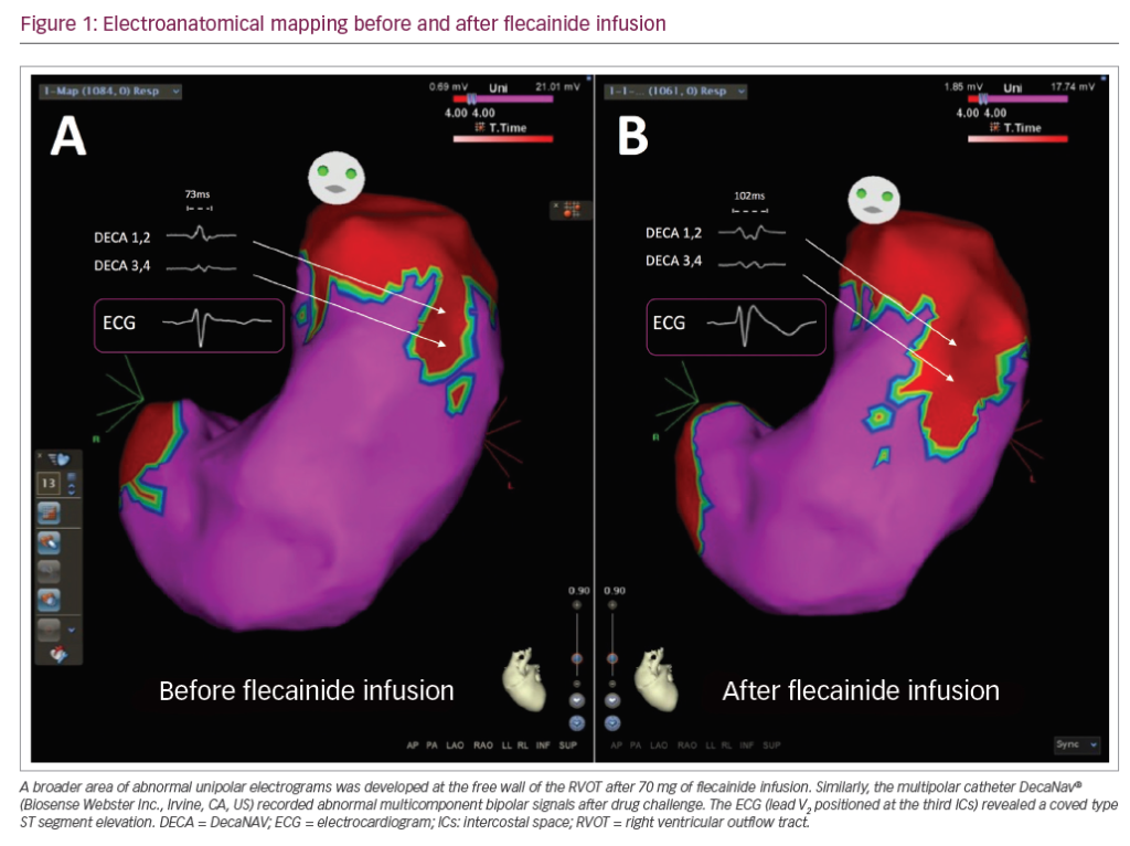
Brugada syndrome (BrS) is a genetic arrhythmia syndrome with increased risk of sudden cardiac death. Unmasking a type I BrS electrocardiogram (ECG) pattern after administration of sodium channel blockers may be associated with an increase in epicardial substrate abnormalities. We ...
Latest articles videos and clinical updates - straight to your inbox
Log into your Touch Account
Earn and track your CME credits on the go, save articles for later, and follow the latest congress coverage.
Register now for FREE Access
Register for free to hear about the latest expert-led education, peer-reviewed articles, conference highlights, and innovative CME activities.
Sign up with an Email
Or use a Social Account.
This Functionality is for
Members Only
Explore the latest in medical education and stay current in your field. Create a free account to track your learning.

