Background: Periodic breathing pattern is a poor prognostic factor of advanced systolic heart failure (HF). It can manifest as exercise-induced oscillatory ventilation (EOV) on cardio-pulmonary exercise testing (CPET); a result of impaired homeostasis. Atrial fibrillation (AF) is also associated with exertional dyspnoea and EOV has not been described in patients with AF and HF.
Objective: To report the prevalence and characteristics of EOV in patients with AF and HF, and the impact of catheter ablation (CA).
Methods: Patients with persistent AF and HF undergoing first-time CA underwent CPET at baseline and 6 months after restoration of sinus rhythm as part of enrolment in the AFHF study (ClinicalTrials.gov identifier: NCT04987723) between January to December 2022. CPET was performed on a semi-recumbent cycle ergometer with minute ventilation, oxygen saturation and carbon dioxide production monitored throughout. A 3-minute warm-up was followed by exercise with the work rate incremented by 10–20 Watts/minute till exhaustion, aiming for 8–10 minutes of exercise and a respiratory exchange ratio of >1.0. EOV pattern was quantified as the average amplitude of oscillation in minute ventilation (Ve) and the percentage duration of oscillatory breathing pattern relative to the total test period (EOV fraction). PROMs were evaluated through contemporaneous completion of the Atrial Fibrillation effect on Quality of Life (AFeQT) questionnaire (rescaled to /100). Paired statistical testing (paired Student’s t-test for parametric and Wilcoxon signed-rank test for non-parametric variables) will be used to compare changes over time.
Results: Of the 17 patients who completed follow-up within the study duration, the mean age was 62 ± 10 years and the average BMI was 30.8 ± 6.2 kg/m2, with 14 (82%) of the cohort male. Significant improvements were seen in peak VO2 (1,473 ± 640 mL versus 1,780 ± 590 mL, p=0.02).
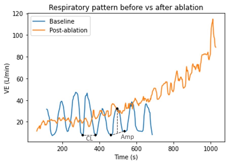
EOV phenomenon was observed in 16 out of the 17 patients (94%) at baseline with an oscillatory amplitude of 10.0 L/min (7.5, 12.2) and a cycle length of 30.2 seconds (23.3, 37.5). Post-ablation, the EOV fraction reduced significantly in 15 (94%) patients (15% [5, 19] versus 6% [4, 12], p=0.01). The oscillatory amplitude (6.9 L/min [4.4, 9.1], p=0.02) and cycle length (25.0 s [18.0, 30.0], p=0.02) also decreased significantly. In line, the symptom severity score improved significantly (47 [28,61] versus 94 [85, 96] p=0.03).
Conclusion: EOV is commonly observed in patients with AF and HF, but significantly improves after CA. This suggests a dynamic nature of the underlying mechanism with potential pathway remodelling in this patient cohort when sinus rhythm is restored. Further research is warranted to better elucidate the relationship between EOV and exertional dyspnoea. ❑
Figure 1: Respiratory pattern before versus after ablation


