
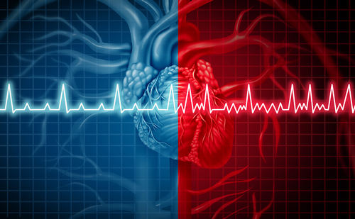
Background: Detection of atrial fibrillation after stroke may be challenging and often requires prolonged monitoring. Atrial fibrillation is known to be associated with imaging markers of atrial structural remodelling such as left atrial dilatation and fibrosis. However, assessment of atrial morphology is challenging with previous approaches lacking standardisation.
Objective: To determine if left atrial morphological parameters assessed using computed tomography with a standardised semi-automatic algorithm are associated with a history of stroke and could be a novel tool to guide therapeutic decisions in patients following stroke.
Methods: Data from the SCOT-HEART trial cohort were retrospectively analysed including 71 patients with a history of stroke, 71 control patients with no stroke or atrial fibrillation and 27 patients with atrial fibrillation. A systematic method of segmentation and morphological assessment was used to derive left atrial volume, shape, wall thickness, mass, sphericity and appendage morphology.
Results: There were no differences in median left atrial volume, left atrial appendage volume or left atrial sphericity between the three groups. Median wall thickness in the Stroke Group, Control Group and AF Group were 1.71 ± 0.51 mm, 1.54 ± 0.45 mm and 1.69 ± 0.55 mm, respectively (p=0.184). There were no differences in the frequencies of different appendage morphologies between groups (p=0.191). On multivariate analysis, current smoking status and CHA2DS2VaSc score were predictive of stroke history but the addition of atrial morphological parameters had little incremental impact on the predictive model for stroke.
Conclusions: Patients with stroke or atrial fibrillation have no major differences in atrial morphological parameters suggesting that these characteristics are not major determinants of clinical disease.
108/Relationship between left atrial wall thickness left atrial morphology and stroke history in the SCOT-HEART trial
Article
Further Resources

Trending Topic
Ventricular fibrillation (VF) is characterized by rapid (>300 beats a per minute), irregular electrical activation with variable electrocardiographic waveforms that prevents coordinated myocardial contraction, resulting in immediate loss of cardiac output.1 It most commonly occurs in the context of coronary artery disease.2,3 Resuscitation efforts are critically time-dependent: with each minute of untreated VF, the survival rate declines […]
Ventricular fibrillation (VF) is characterized by rapid (>300 beats a per minute), irregular electrical activation with variable electrocardiographic waveforms that prevents coordinated myocardial contraction, resulting in immediate loss of cardiac output.1 It most commonly occurs in the context of coronary ...
The official abstracts from the Heart Rhythm Congress (HRC) 2025

It is with pride and gratitude that we reflect on the remarkable 10-year journey of European Journal of Arrhythmia & Electrophysiology. With the vital contributions of all of our esteemed authors, reviewers and editorial board members, the journal has served ...

Postural tachycardia syndrome (PoTS) is a chronic condition, predominately affecting women between the ages of 15 and 50, and prevalence is estimated to be 0.2%.1,2 It is characterised by an abnormal response by the autonomic nervous system to changes in posture causing lightheadedness ...
Catheter ablation (CA) is an invasive adjunctive therapeutic option for atrial and ventricular arrhythmias, in uncontrolled cases, following optimal medical therapy.1,2 The validation of 3D mapping and its implementation into clinical usage has been a remarkable achievement in the field ...

Welcome to the latest issue of the European Journal of Arrhythmia & Electrophysiology, the official journal of the Arrhythmia Alliance. We are honoured to present a series of compelling articles that reflect cutting-edge developments and diverse perspectives in this ever-evolving ...

Ventricular tachycardia (VT) ablation presents a challenging and growing area of focus in the realm of complex ablation, with promises of improved patient’s quality of life and clinical outcomes.1,2 Classic approaches to VT ablation centered on arrhythmia induction and ...

For decades, right ventricular pacing (RVP) has been the leading pacing technique and has been proven to be effective in treating patients with symptomatic bradycardia. However, dyssynchrony caused by non-physiological ventricular activation results in pacing-induced cardiomyopathy occurs in approximately 15% of ...

Supraventricular tachycardia (SVT) is an important cause of intrusive palpitation symptoms. This expert interview explores a nurse-led end-to-end pathway for the diagnosis, assessment and management of this condition. It was conducted at the Barts Heart Centre of Barts Health NHS ...

Highlights Failure to target and deliver durable radiofrequency lesions to all functionally critical areas has been identified as an important cause of ventricular tachycardia recurrence and is particularly challenging in deep midmyocardial or very extensive substrate where point-by-point radiofrequency ablation ...

Bradycardia, renal failure, atrioventricular nodal blockade, shock and hyperkalemia (BRASH) syndrome is a rare, often unrecognized but potentially life-threatening clinical entity that is characterized by the pentad of symptoms after which it is named. BRASH syndrome is seen most commonly ...

Radiofrequency (RF) ablation involves delivering medium-to-high-frequency alternating current, which is electrically conducted to cardiac tissue that constitutes a part of the circuit. The unipolar circuitry contains the generator, transmission lines, catheter, cardiac tissue, interposed tissue and dispersive electrode (ground pad/...
Log into your Touch Account
Keep track of your clinical interests and newsletter subscriptions.
Sign up with an Email
Or use a .
Register now for FREE access
Already registered? Login below.
